Dental Disease in Rabbits
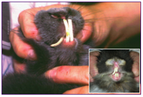
Rabbit with severely overgrown incisors.
Inset: Rabbit with single overgrown incisor.
Today, dental disease in rabbits is a very commonly diagnosed problem and is now recognized as the underlying cause of numerous other disorders. For this reason an understanding of dental health and physiology are crucial for veterinarians that treat rabbits and for rabbit owners alike.
Rabbits are herbivorous, naturally eating a wide variety of vegetation and roughage. A unique feature of rabbits is that all of their teeth are open rooted which means that their teeth grow continually throughout their life. A healthy rabbit eating a proper diet constantly chewing will wear down the teeth as they grow.
Signs of dental disease
Signs of dental disease are very broad and may be non specific. Early signs can be subtle and may not be immediately noticeable to the rabbit owner. The rabbit may change its food preferences, stop eating certain things that may be difficult to chew, or drop food from its mouth. The rabbit may have some weight loss or may look unkempt from a change in grooming habits, especially if the incisors are overgrown. Advanced signs of dental disease may be excessive salivation, loss of appetite, malodorous breath, and severe weight loss. Also, the rabbit may present for another problem such as GI stasis, an abscess, or an infected tear duct, all secondary to the primary dental disease.
Causes of dental disease
The most common causes of dental disease in rabbits are genetic and dietary. Genetic predisposition is very common. Unfortunately with inbreeding, some rabbits are born with malocclusion. This is commonly seen with the incisor teeth which overgrow and curl around when they do not meet properly. These rabbits often need to have their teeth trimmed every 1-2 months or have the incisor teeth extracted. Some rabbits are born with elongated skulls which also leads to dental occlusion problems. Another common cause is diet. Many rabbits are primarily fed a diet of pellets. Because the pelleted food is dense in nutrient content and is already pulverized, the rabbit chews less. Rabbits’ molars curl as they continually grow. With decreased wear from less chewing, the lower molars develop points that grow into the tongue and the upper molars develop points that grow out into the cheeks. These changes occur gradually with time and can lead to many secondary problems including abrupt loss of appetite and infections. Acquired dental disease with deterioration of the tooth quality, malocclusion and elongation of the roots with periapical abscesses is another form of dental disease in rabbits. Metabolic bone disease, genetics, and diet are all proposed causes for this problem.
Diagnosis
Because dental disease is so important in rabbits, a dental exam should be a part of the annual physical examination. As part of the exam, the veterinarian will check for the overall body condition, the condition of the coat, drainage from the eyes, swelling under the jaw bone, and saliva staining of the fur under the chin, which can all be indications of dental disease. The incisor teeth are checked by lifting the lips to check their length and occlusion. The sides of the cheeks are palpated for any sharp protrusions. The veterinarian will insert a speculum and/or an otoscope to visually evaluate the molars. Very often the veterinarian is able to see points or spurs on the molars and will recommend a more complete dental exam and filing of the molars under anesthesia based upon his/her observations.
The dental examination under anesthesia can be performed safely after a preanesthetic blood profile and full exam are performed to be sure that the rabbit is in good health and low risk for anesthesia. The anesthetic agents are chosen based upon the age and health status of the rabbit, the length of time of anesthesia and the expected difficulty. Pain medications are also utilized to decrease the stress of the procedure and improve the rabbit’s recovery. Specialized instruments are utilized to visualize the oral cavity including cheek spreaders, an incisor speculum, and a special light source. Special diamond files, rongeurs, and straight dental burs on a low speed drill are used to remove points and file the teeth down. In recent years a system for grading the extent of dental disease based on radiographs has been developed. While under anesthesia, the x-rays may be taken to evaluate the tooth roots for elongation and periapical abscesses.
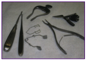
Instruments used in rabbit dentistry
Treatment
Treatment of dental disease will depend on the stage of the disease at the time of diagnosis. In all cases, rabbits that are severely debilitated from anorexia, GI stasis, or severe infection will need to be stabilized first. Administration of fluids, maintenance of body temperature, pain medications and antibiotics may be the first steps along with restoring adequate nutrition. Once the rabbit is stable, it may safely undergo anesthesia for the dental exam.
For severely overgrown incisors, a dental drill can be used to trim the incisors. This is a problem that tends to be chronic and repeat trimmings at regular intervals are needed. For some rabbits the best option is to extract the incisors. Incisor extraction is a major surgical procedure and is done under anesthesia. The two long upper and two long lower incisors and the two upper peg teeth are extracted with special elevators. The rabbits are sent home on antibiotics, pain medications and some soft food for a few days. Fortunately the rabbits are able to prehend food with their lips and do well with this procedure after a few days of nursing care.
Blocked tear ducts are another problem seen in rabbits with overgrown incisor roots. The tip of the root is very close to the tear duct so inflammation or elongation of the root can cause a blockage. The owner will notice tears and sometimes pus spilling onto the face from the eyes. To diagnose this problem an x-ray contrast medium can be instilled into the duct and an x-ray taken to demonstrate the blockage. Another test is to place fluorescein stain solution into the eye and check with a UV light for the dye passing out of the nostril. This is a difficult problem to treat. Sometimes, the tear duct can be flushed while the rabbit is awake or under anesthesia and the drainage can be restored. Repeated flushing may be needed. Often the veterinarian will prescribe an antibiotic ointment to help prevent an infection. The owner should keep the area under the eye clean and dry to avoid secondary infection.
For severely overgrown molars or molars with points, a low speed dental drill with a straight dental bur can be used to file down the molars and correctly reshape the teeth. Periapical infections of the molar roots are common in acquired dental disease and often lead to the formation of abscesses and infection in the surrounding bone. If an abscess is present, surgical treatment is required to remove the entire abscess and the capsule. Involved bony tissue should be debrided. Antibiotic impregnated beads or antibiotic saturated umbilical tape drains can be placed into the abscess cavity. Sometimes intraoral extraction of diseased molars is also necessary along with flushing of the root socket.
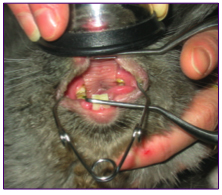
View of rabbit’s mouth during dental procedure.
Prevention
Unfortunately most dental diseases in rabbits tend to be chronic. Exams and filing need to be done on a regular basis and infections treated promptly. The best way to prevent dental disease is to make sure the rabbit is on a proper diet with good quality hay and leafy greens offered every day. Avoid feeding mainly pellets. Be aware of the many signs of dental disease and call the veterinarian promptly if a problem is suspected. Regular yearly physicals with a dental exam are essential and allow early diagnosis of a problem and the best prognosis for resolution.
©2006 by Alexandra Kilgore, DVM
Dr. Alexandra Kilgore is a veterinarian at Littleton Animal Hospital in Littleton, MA.